
research activities in broadband infrared diagnostics
— Mihaela Zigman —
Exploring novel routes to early cancer detection with femto- and attosecond metrologies
Cancer ensues when a single cell acquires the capacity for uncontrolled reproduction, proliferates aggressively and invades other tissues.
Increased sensitivity and fidelity of early cancer detection, and a better quantitative understanding of the initial changes that drive tumor growth at the molecular level are required to advance current medical anti-cancer strategies.

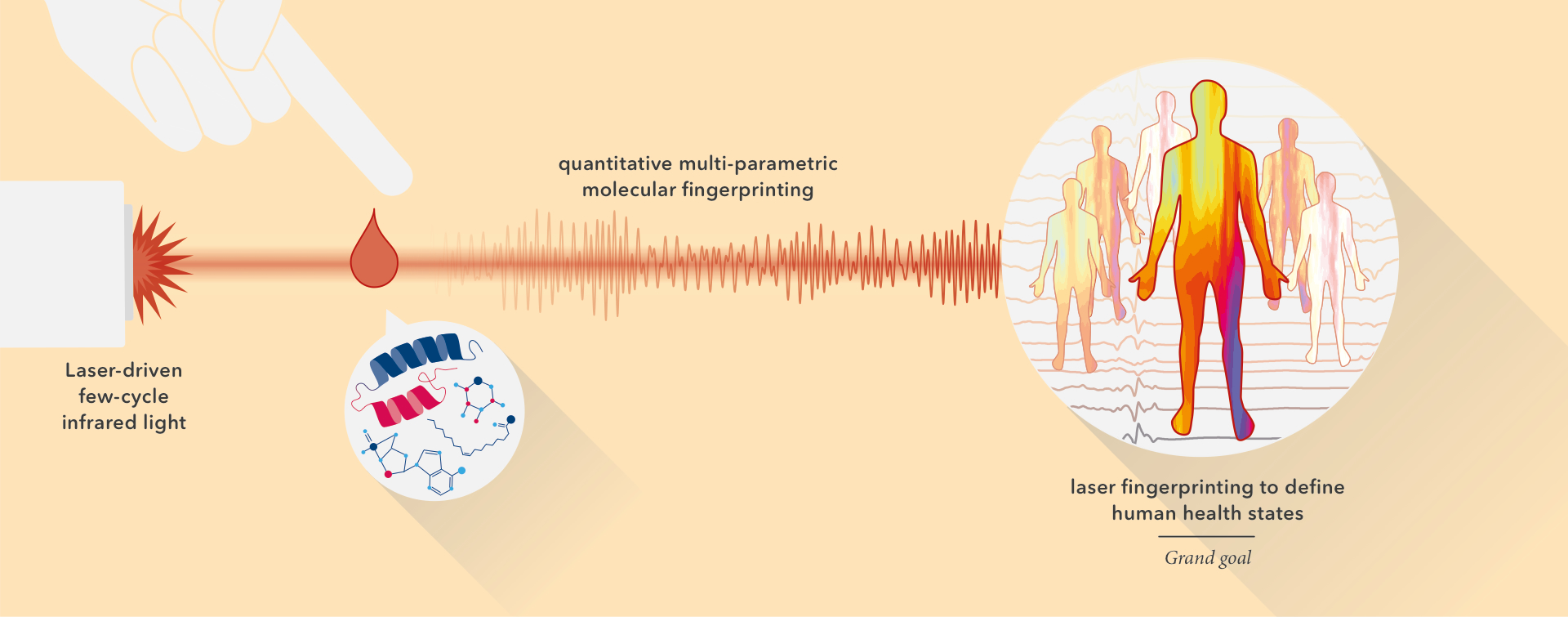
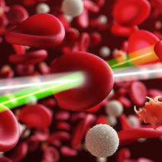
To this end, we are exploring the application of molecular spectroscopy based on a powerful broadband femtosecond infrared laser source, together with time-domain metrology for vibrational spectroscopy to cancer diagnosis.
Our laser-based vibrational infrared spectroscopy set-up (developed by the teams of Dr. Pronin and Dr. Pupeza) combined with Raman spectroscopy (developed by the team of Dr. Fattahi) provide unprecedented capacity to acquire complete amplitude and phase information for biological samples.
This will allow us to detect very early cancerous changes — by quantitatively detecting changes in characteristic absorptional fingerprints of biomolecules and of metabolites originating from living human cells.

Together, the ability to quantifying pathological pre-malignant changes and their spatio-temporal dynamics fundamental to cancer biology will enable to:
1.) to functionally interrogate the mechanisms how specific genetic alterations trigger cancer in real-time in vivo and 2.) to provide robust molecular, cellular and metabolic snapshots reveal the earliest pathological transitions in the human body.
It is this unique combination of laser-based technologies and biological paradigms that promise to extend our understanding of fundamental principles in cancer cell biology. In the future, we expect our approach to have an impact on clinically relevant methods for cancer detection and therapy monitoring in patients.

Graphical Abstract
learn more about broadband infrared diagnostics
— Mihaela Zigman —
Tools, Techniques & Labs
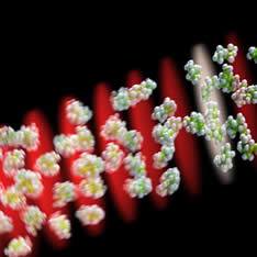
Molecular vibrational infrared laser spectroscopy, by means of femtosecond infrared absorption spectroscopy and stimulated Raman scattering, takes advantage of the fact that each molecular species exhibits a characteristic set of vibrational frequencies that depends on its atomic structure. The complete set of these frequencies, most of which lie in the infrared spectral range, constitutes a unique »molecular fingerprint«.
We exploit of the potential of absorption spectroscopy to acquire molecular fingerprints of biological samples that contain highly complex yet state-specific mixtures of organic molecules.
A further advantage of our approach is the ability to probe biological samples both in liquid phase (human cells, blood serum) and gaseous phase (small volatile compounds, human breath).
Our technology operates in a label-free manner, thereby obviating the need for complex biochemical sample preparation. Finally, we are devising new approaches to data analysis, which will facilitate the application of broadband infrared detection to high-throughput screening of large sample sets.
-
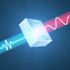
 Molecular spectral fingerprint measured over time with field-resolved spectroscopy (x axis time, y axis spectral wavenumbers, the colors encode spectral intensity).
 -
-
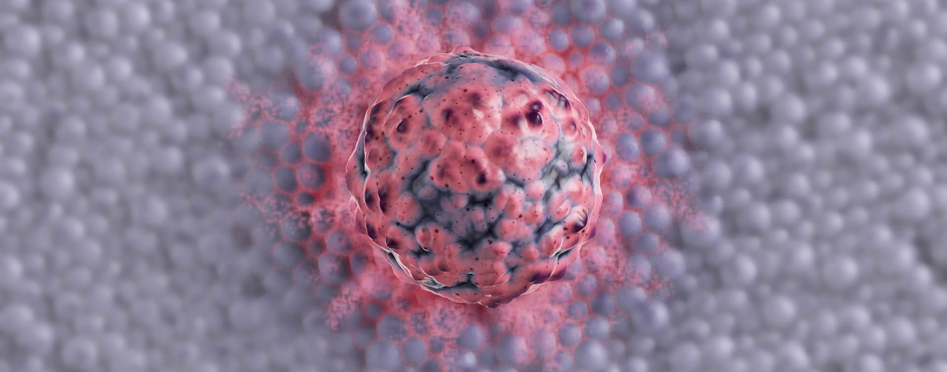
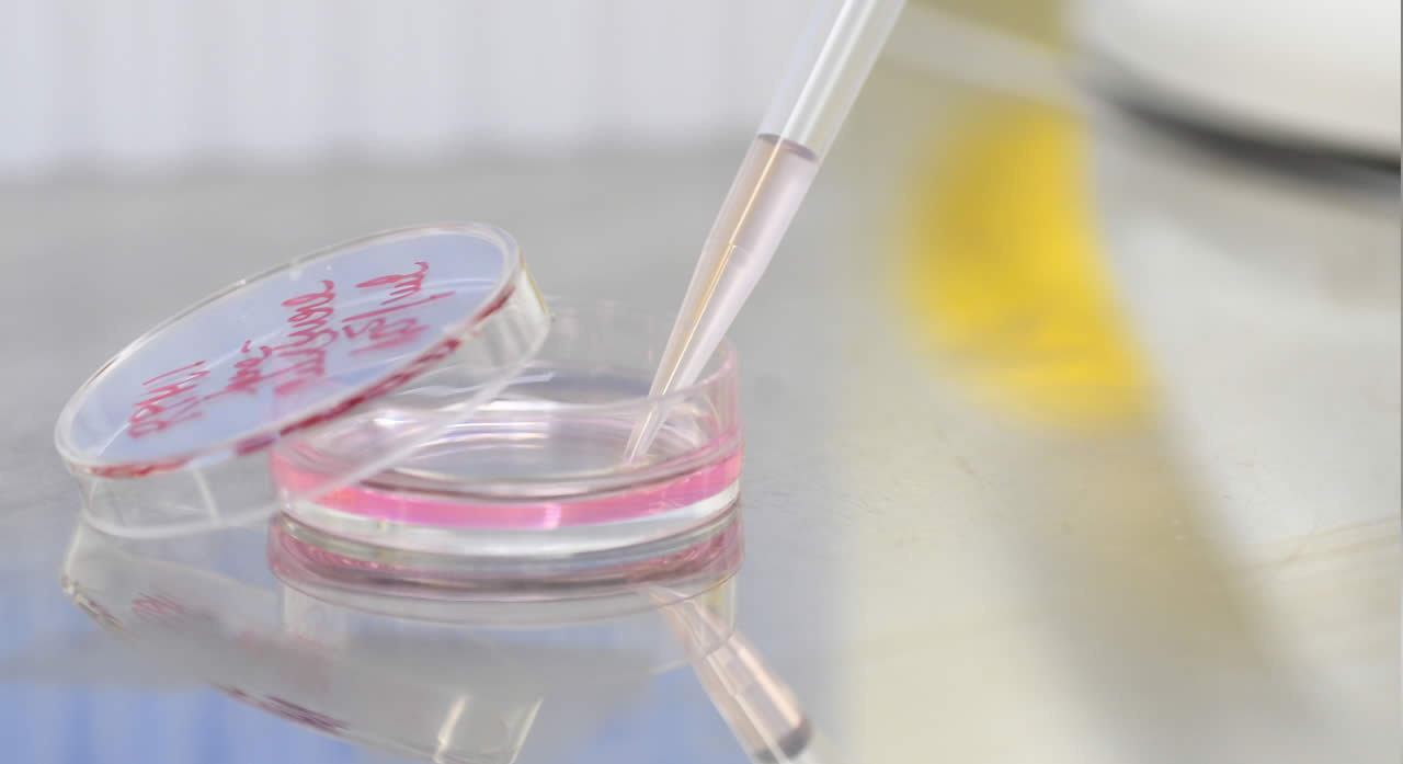 Living human cells cultured in a petri dish.
 -
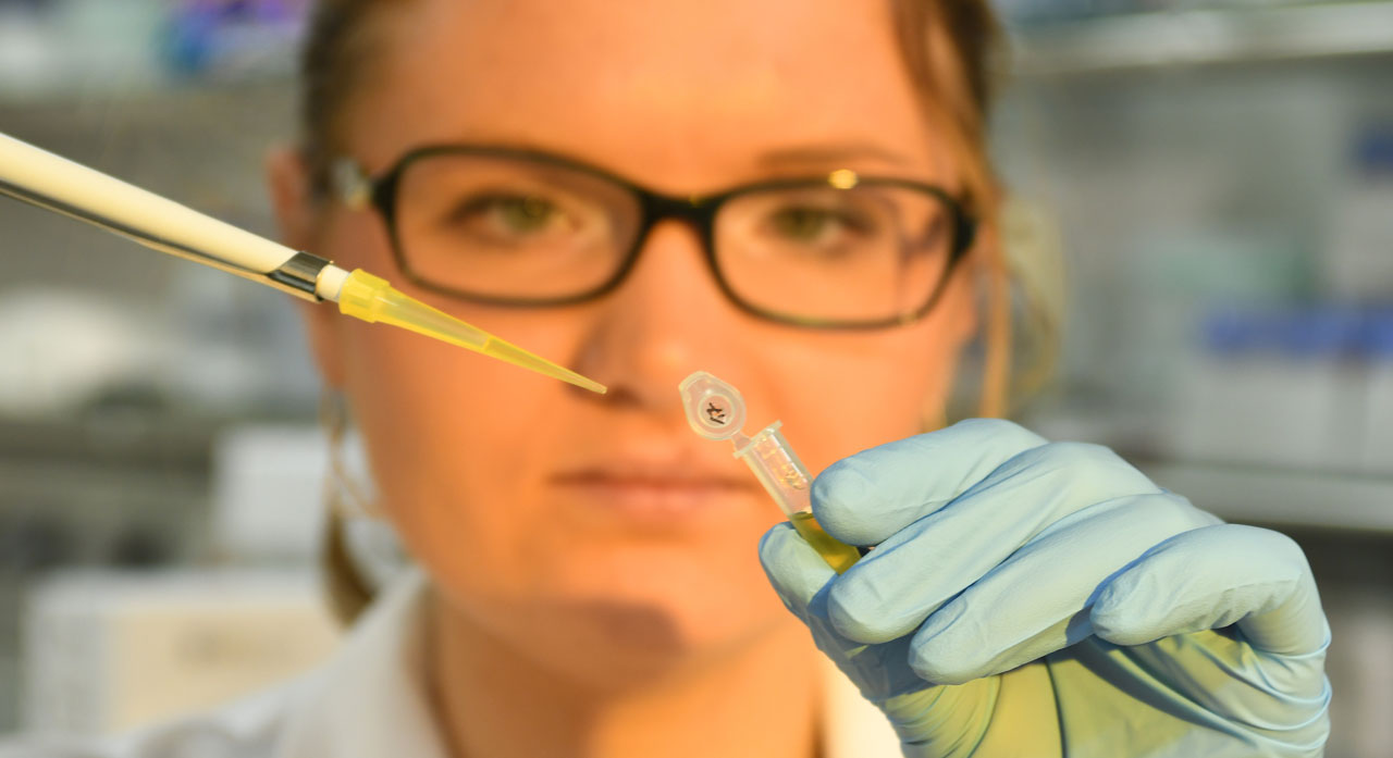
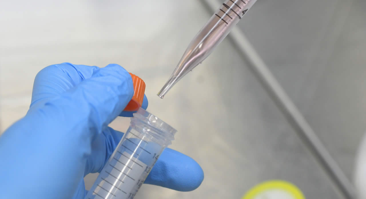 Molecular biology lab.
 -
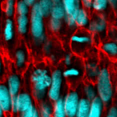
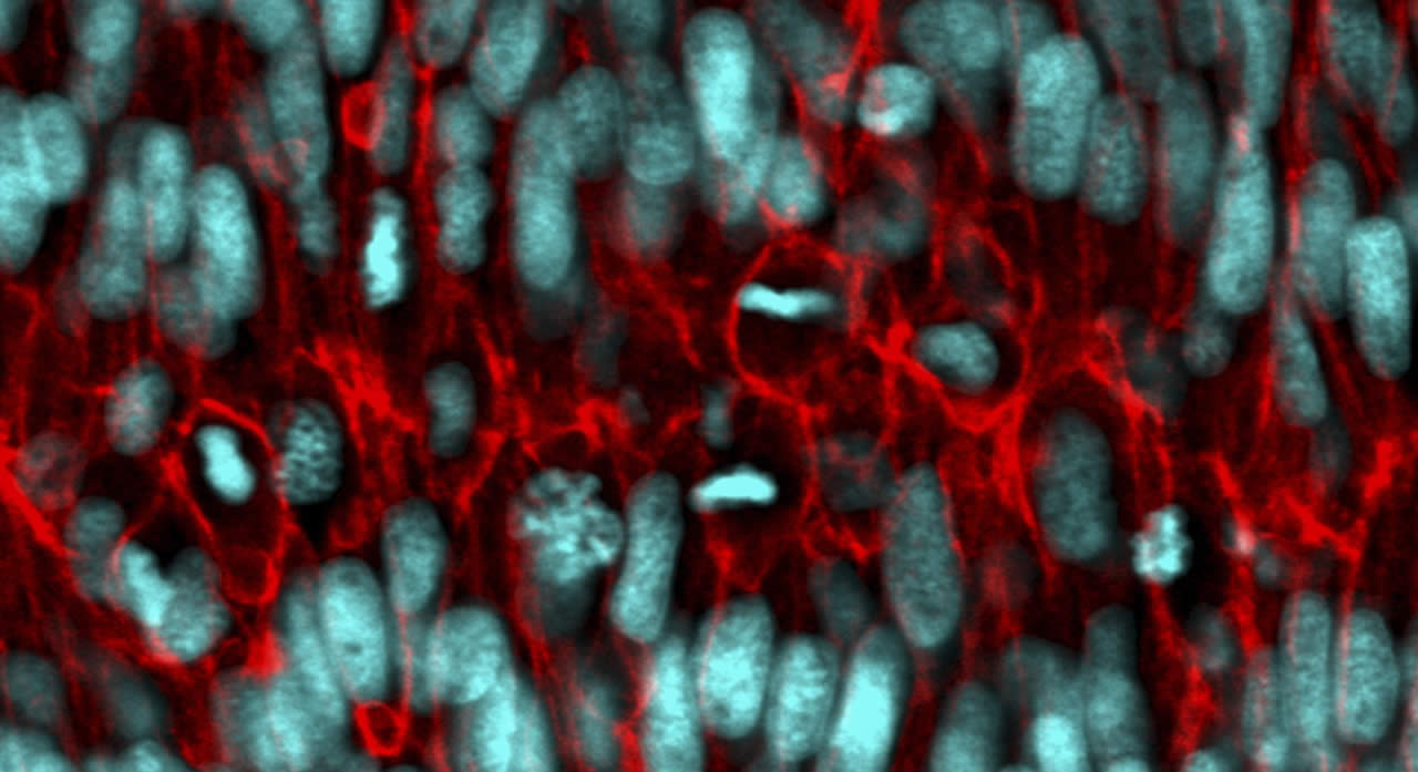 Actively growing and dividing living cells deep within an intact organ (laser scanning confocal micrograph of a double transgene: cell nuclei marked in cyan, cell membranes marked in red).
In collaboration with Dr. Oleg Pronin, Dr. Ioachim Pupeza, Dr. Hanieh Fattahi and their teams, we are setting up a state-of-the-art femtosecond lab infrastructure to pursue the above goals outlined above. Our infrastructure encompasses a molecular biology lab, state-of-the-art manual and semi-automated Fourier-transform infrared spectrometers (FTIR) for benchmarking, and several broadband femtosecond-based spectroscopic measurement systems for femtosecond infrared absorption spectroscopy and stimulated Raman scattering.